In 1923, some studies discovered that humans glow at visible frequencies when the light source is strong enough. The truth is that from the time we are in our mother's womb until we leave this world, we actually glow.
This may be a controversial topic, but if we can detect these 'biophotons', we could potentially learn more about what's going on under our skin.
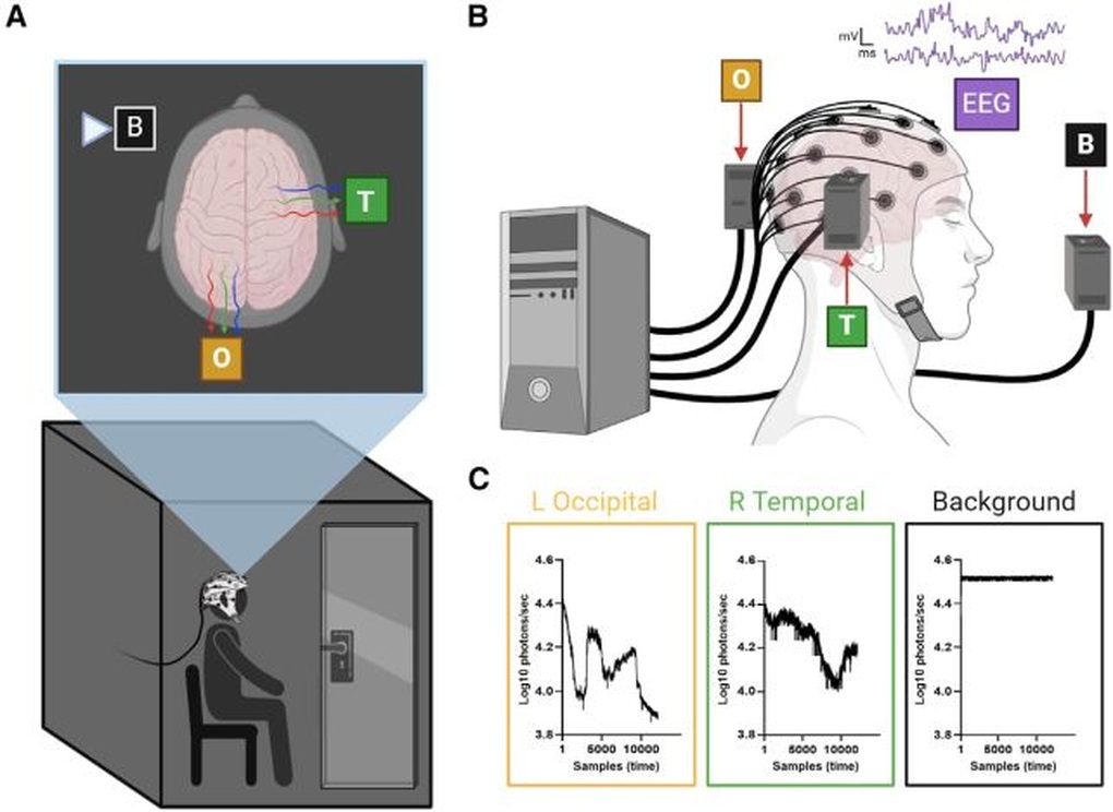
In a new study, a team of researchers led by biologist Hayley Casey at Algoma University in Canada investigated the extremely faint glow of a specific mass of tissue, the brain, located inside the skulls of all living people.
The team carefully recorded the faint glow of the human brain from outside the skull and found that it changed depending on the brain’s activity at any given moment. This opened up a new possibility for assessing brain health: a previously undeveloped technique that scientists call photoencephalography.
"To provide the first evidence that ultraweak photon emission (UPE) from the human brain can be used as a functional state monitoring data, we measured and characterized the number of photons on the heads of participants while they were at rest or during auditory activity," the study report reads.
The team demonstrated that the UPE signals originating from the brain differed from background photon measurements. In addition, the study results showed that when performing certain tasks, the number of UPEs emitted was at a certain specific level.
Everything in the universe that has a temperature above absolute zero, including humans, emits a type of infrared radiation called thermal radiation. When we talk about UPE, it is a separate phenomenon distinct from thermal radiation.
UPE is emitted at wavelength ranges close to visible light and is the result of electrons emitting photons as they lose energy, a normal byproduct of metabolism.
The team sought to clearly distinguish UPEs in the brain from background radiation and determine whether these UPEs appeared at levels corresponding to different brain activities.
They placed each study participant in a darkened room. The participant wore an electroencephalogram (EEG) cap to monitor brain activity, and photomultiplier tubes were positioned around them to record any light emissions. These vacuum tubes are extremely sensitive, able to detect even the faintest light.
The results show that not only is UPE real and measurable, but there is also a clear correlation between emitted UPE and each different activity.

The researchers say future research may look into how neuroanatomy might influence UPE output, as well as how different activities manifest in UPE models, rather than just the two resting and active brain states.
They also said that it is currently not possible to confirm whether each individual has a unique UPE similar to fingerprints. This is also a topic that scientists are interested in studying.
Source: https://dantri.com.vn/khoa-hoc/nao-phat-ra-anh-sang-bi-mat-ma-ban-khong-he-biet-20250619022639708.htm


![[Photo] Party and State leaders visit President Ho Chi Minh's Mausoleum and offer incense to commemorate Heroes and Martyrs](https://vphoto.vietnam.vn/thumb/402x226/vietnam/resource/IMAGE/2025/8/17/ca4f4b61522f4945b3715b12ee1ac46c)


Comment (0)